Ultrasound Services
Every exam is performed by registered, highly experienced technologists and read by board certified physicians. If you do not see the exam you need, call us at (314) 782-2034 and we will help.
Abdominal & Pelvic
Abdominal Complete
Liver, gallbladder, bile ducts, pancreas, kidneys, spleen, aorta
Abdominal Limited RUQ
Liver, gallbladder, pancreas, bile ducts, right kidney
Aorta
Abdominal aortic aneurysm evaluation
Aorta Screening
One screening exam per lifetime
Pelvis Complete & Transvaginal
Pelvis Limited
Urinary bladder (male)
Renal
Kidneys, ureters, and bladder
Renal with Doppler
Renal artery and vein stenosis, thrombosis
Retroperitoneal Limited
One structure or part of a structure: aorta, IVC, kidney
Hernia Evaluation
Pregnancy & GYN
OB Ultrasound
Early pregnancy through term
Level 1 OB Anatomy Scan
Standard first, second, or third trimester
Transabdominal & Transvaginal Pelvic Ultrasound
Follicle Monitoring
Vascular
Venous Doppler (DVT check)
Upper & lower extremity, unilateral or bilateral, same day with STAT interpretation
Arterial Doppler & ABI
Upper & lower extremity, ankle brachial indices
Carotid Doppler
Renal Artery Duplex
Aorta Duplex
Abdominal Doppler
Aorta, IVC, mesenteric, renal, liver, spleen
Venous Insufficiency Studies
Temporal Artery Duplex
Heart
Transthoracic Echocardiogram
With and without Doppler
Musculoskeletal
Extremity Complete
Shoulder, elbow, wrist, hand, finger, hip, knee, ankle, foot, toes, including arthritis studies
Extremity Limited
Soft tissue masses or a localized area, no joint included
Tendons, Ligaments & Muscles
Superficial Nerves
Mass Characterization
Dynamic Imaging
Structures viewed in motion, which MRI cannot show
Hand & Foot Ultrasound
For rheumatology evaluation
Thyroid & Soft Tissue
Thyroid Ultrasound
Soft Tissue Head & Neck
Parathyroid, submandibular, scalp, soft tissue masses
Chest & Back Soft Tissue
Soft tissue masses, lipomas
Soft Tissue Body Masses
Scrotum / Testicular
Masses, hernia, pain
Scrotum / Testicular with Doppler
Torsion, varicocele, AVM
Exams performed here
A selection of studies performed at Ultrasound First, captured on our own systems. Some of these findings are difficult to see on any other imaging, and some require positioning and patience that most facilities will not take the time for.
Bicuspid aortic valvemoving image
An echocardiogram recorded in real time. This valve opens with two leaflets instead of the usual three.
Deep vein thrombosismoving image
A clot inside a leg vein, filmed as it moves. Exams like this are read the same day with results called straight to your doctor.
Patent foramen ovalemoving image
Color Doppler showing blood crossing an opening between the upper chambers of the heart.
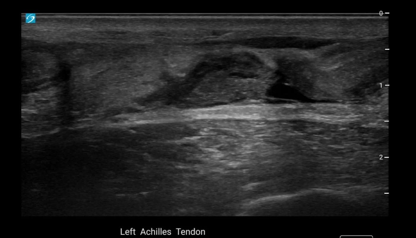
Achilles tendon tear
The gap where the tendon has pulled apart, visible without radiation or a scanner tunnel.
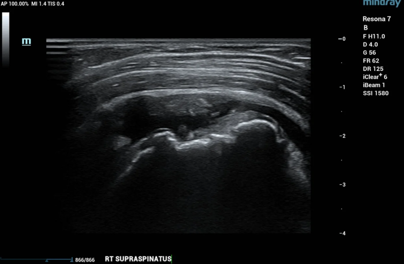
Full thickness rotator cuff tear
A complete tear of the supraspinatus tendon in the shoulder, one of the most common causes of shoulder pain.
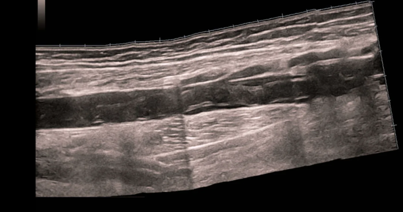
Clot along the femoral vein
Thrombus filling the length of a thigh vein, found on a same day venous Doppler exam.
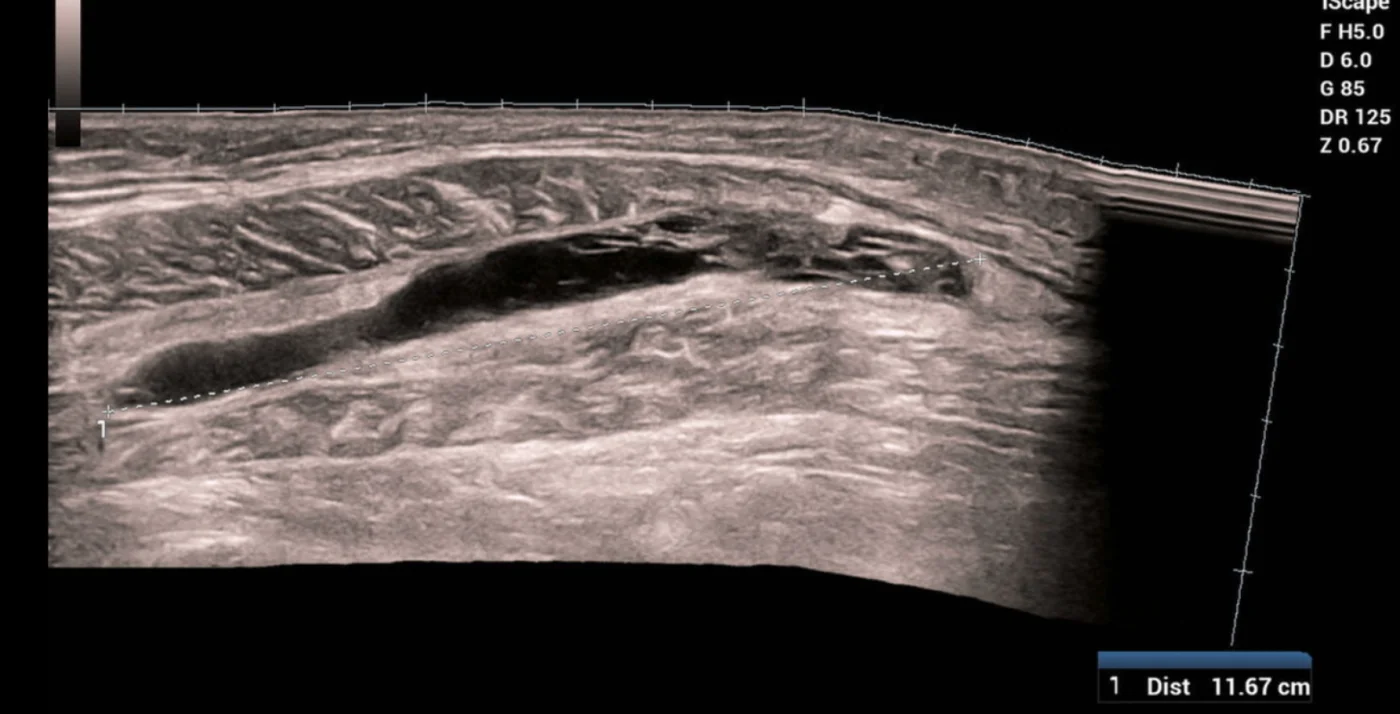
Calf muscle bleed
An intramuscular hematoma in the gastrocnemius, a frequent cause of sudden calf pain after a strain.
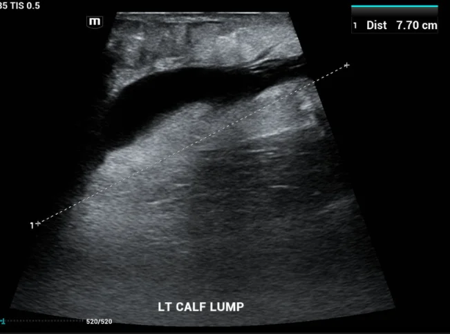
Morel-Lavallee lesion
Fluid collecting between tissue layers in the calf after an injury, measured here at 7.7 cm.
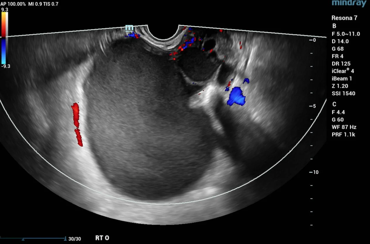
Ovarian endometrioma
A cyst on the ovary seen with color Doppler during a pelvic exam.
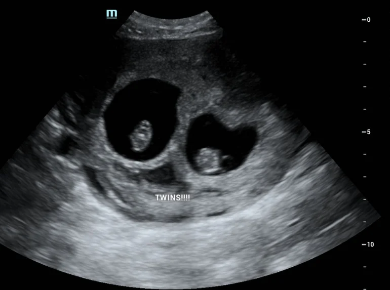
Twins
Two gestational sacs on an early pregnancy exam.
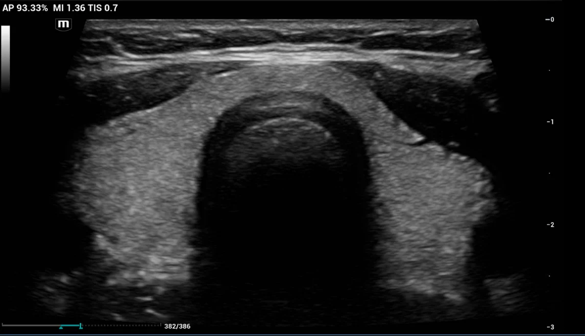
Normal thyroid
A healthy thyroid gland. Knowing what normal looks like is half of what makes a finding meaningful.
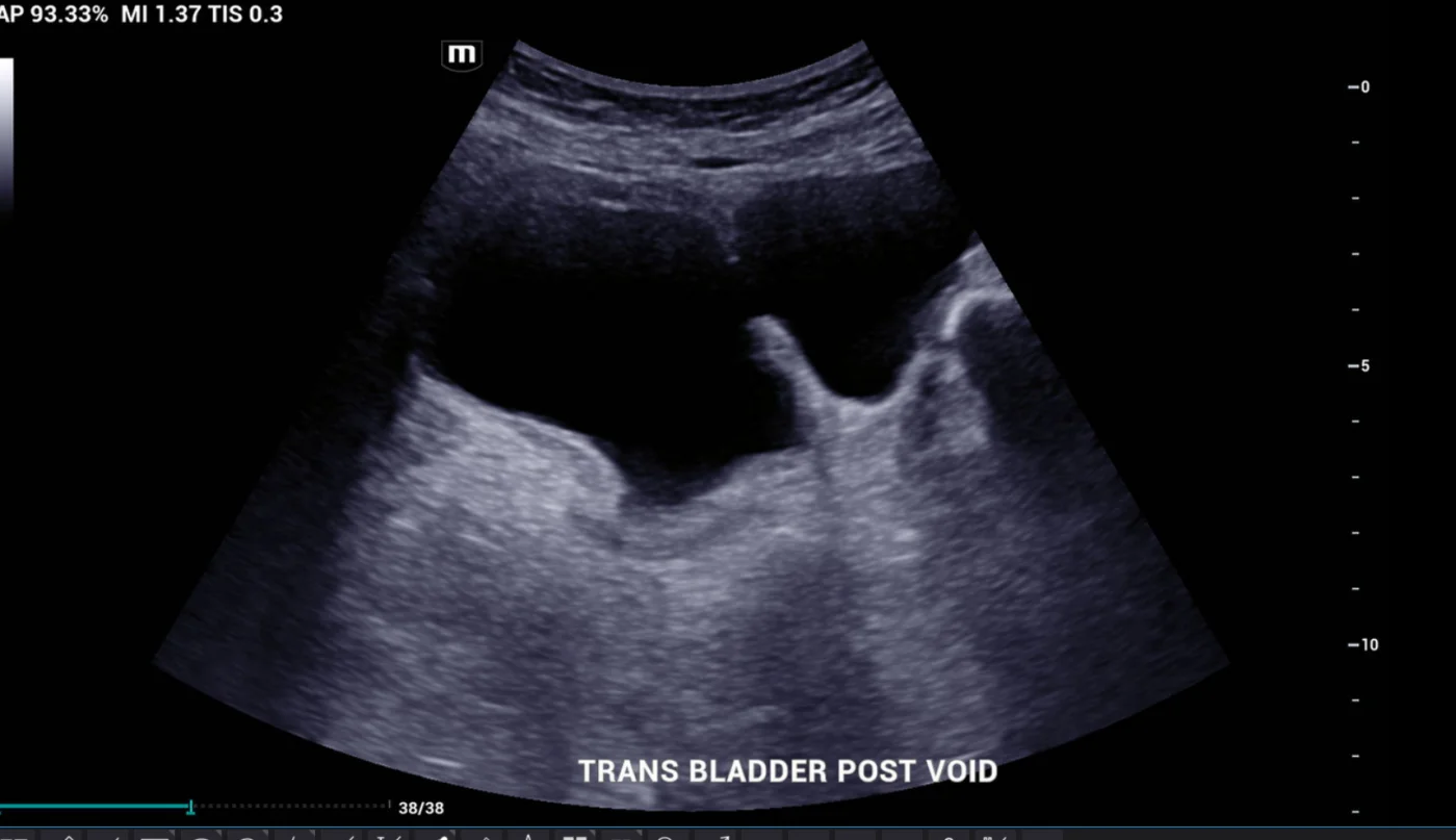
Bladder diverticulum
An outpouching of the bladder wall, caught on a post void image during a pelvic study.
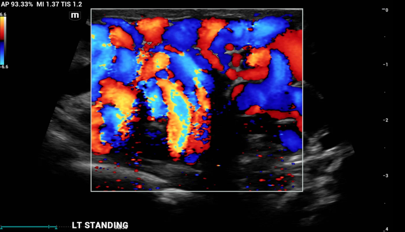
Scrotal varicocele
Enlarged veins imaged standing, a position many facilities will not take the time to scan.
All images and clips were captured on our own equipment and contain no patient information.
Common questions
Do I need a doctor’s order for an ultrasound exam?
Yes, diagnostic exams are performed with an order from your doctor. Orders can be faxed to (314) 931-3130, and we are glad to help coordinate with your physician.
Can you check for blood clots the same day?
Yes. Venous Doppler DVT exams for the upper and lower extremities are available same day with STAT interpretation.
What if I do not see the exam I need listed?
Call us at (314) 782-2034. Our team offers a comprehensive range of diagnostic ultrasound exams and will help you find what you need.
How much do exams cost?
Most exams cost less than $300, far less than hospital and imaging center fees. We accept most major insurance, workers compensation, and self-pay.